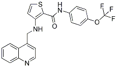
The aim of this study was to probe if HDAC inhibition has any effect on human microvascular BMS-907351 pericytes with regards to proliferation, cell viability, migration and differentiation into profibrotic connective tissue cells in vitro. Furthermore, given the growing importance of the role of the pericyte in the regulation of the vasculature and the Talazoparib process of angiogenesis focus was directed towards potential modifications of expression patterns of mRNAs coding for proteins known to be involved in the angiogenic process in pericytes in response to HDAC inhibition. The effect of HDAC inhibition on pericyte proliferation was investigated. Inhibition of pericyte proliferation was seen already at a concentration of 1 mM VPA. Although the VPA analogue Penta which lacks HDAC inhibitory effects had a statistically significant effect on pericyte proliferation the effect did not approximate that observed in pericytes exposed to VPA suggesting that the anti-proliferative effect was to a large extent  due to VPAs ability to inhibit HDACs. However, a certain effect on pericyte proliferation due to the “off target effects” of VPA cannot be excluded. The notion that it is HDAC inhibition that is responsible for the effects seen on pericyte proliferation were further supported by experiments using TSA, another more potent HDAC inhibitor, which had similar anti-proliferative effects on pericytes when compared to VPA. The CFDA-SE incorporation experiments show that VPA, in a concentration dependent manner, limits the expansion of the pericyte population over the course of several cell divisions, and resulted in a higher percentage of pericytes retaining the dye. Previous studies addressing the potential role of HDAC inhibitors in blood vessel biology have focused on the endothelium. These in vitro results show that HDAC inhibition has an anti-proliferative effect also on pericytes and limits the expansion of the pericyte population over time. The present investigation shows that HDAC inhibition perturbs pericyte migration. When analyzing results from migration assays cell proliferation and viability must be taken into account in that a decrease in proliferation/viability may be misinterpreted as an inhibition of migration. In the present investigation no significant changes in the number of viable cells was seen after exposing pericytes to the concentrations of VPA used in the migration assay. Thus, cell death should not influence the observed results. At the 24-hour time point inhibitory effects on proliferation, but not migration, were seen in pericytes exposed to 1 mM VPA. VPAs inhibitory effect on migration was first significant in pericytes exposed to 3 mM VPA. Thus, a decrease in pericyte proliferation should not influence the observed results. The HDAC inhibitor TSA had a similar effect on pericyte migration. No effect on migration was observed when cells were exposed to the VPA analogue Penta suggesting that HDAC inhibition and not “off target effects” of VPA lead to the observed inhibition of pericyte migration. The results show that HDAC inhibition inhibits pericyte migration. The present investigation shows that VPA induces and upregulates expression of mRNAs coding for proteins involved in angiogenesis in pericytes in vitro. HDAC inhibition has been shown to inhibit angiogenesis in vivo and endothelial cell proliferation in vitro. No studies to our knowledge have addressed the role of pericytes in HDAC mediated effects on angiogenesis. Several pathological states in adults are initially characterized by an excessive and aberrant angiogenesis.
Vascular regression and pruning followed by vascular maturation is required for the finalization of tissue reparative
Leave a reply