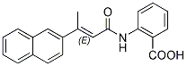
As in previous work, there was little histologic evidence of tissue damage. The statistical parametric maps shown in Figure 6 reveal the time-course and spatial extent of the active neuronal circuitry traced out by Mn2+ induced hyperintensities in the MR images of wildtype and NET KO mice. Table 1 indicates which anatomic structures display Mn2+ induced hyperintensities as a function of time after Mn2+ Dabrafenib citations injection into the PFC. Pathway that is influenced by mesolimbic and mesocortical dopaminergic projections. Similarities and differences between the NET KO reported here and the other two monoamine transporter knockout strains, DAT KO and SERT KO, are displayed in the simplified schematic diagrams summarizing the SPM analyses. These results reveal different time courses of Mn2+ tracing in NET, DAT and SERT KO mice. The SERT KO MEMRI tracing extends more posteriorly than traces from their wildtype littermates. Both the DAT KO and NET KO tracings are truncated compared to those in their wildtype littermates, extending only as posterior as the SNr. The prefrontal cortical and striatal neurons normally project to multiple brain regions, including posterior regions in the midbrain and pons. There is a growing body of LY294002 literature indicating the subtleties, complexities and interrelatedness of the consequences associated with NET, DAT, and SERT deletions. NET KO can up-regulate DAT and SERT in several brain regions. SERT and DAT can accumulate and release NE as a result of heterologous “false transmitter” uptake of the transmitter, although “occult” reuptake is unlikely to account for many effects observed in these mice. There is considerable evidence for adaptations in the other monoamine systems when one transporter is deleted, which is perhaps best exemplified by the unusual rewarding effects of selective NET and SERT blockers in DAT KO mice. The differences in the MEMRI tracing in posterior brain regions may indicate that the balance of activity between anterior and posterior projections from the PFC is altered in these monoamine knockout mice. This change in active connectivity may underlie the characteristic behavioral consequences of each of these knockouts, such as the anxiety-like phenotype of the SERT 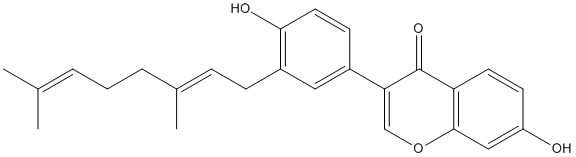 KO, as contrasted with antidepressant-like phenotype of the NET and DAT KO mice. Thus, the mainly frontal MEMRI tracing in DAT and NET KO mice is likely to be indicative of more robust activity and/or connectivity in the frontal portion of the reward circuit of the DAT and NET KO mice compared to wildtype or the SERT KO mice. In the context of understanding the genetic basis of mental illness, Robbins and Arnsten review monoaminergic influences on executive functions of the PFC in rodents, nonhuman primates and humans. They note that much subtler genetic alterations in norepinephrine and dopamine signaling may have significant influences on susceptibility for major mental illnesses. Therefore findings about these mixtures are most often attributed to CUR. CUR belongs to the pharmacopoeia of Asian traditional medicine or alternative medicine to treat inflammatory diseases and a wide range of disorders. Given the role of inflammation in the promotion of chronic human diseases including Alzheimer’s disease, chronic obstructive pulmonary disease, cataract, diabetes, and cancer, CUR has deserved extensive research.
MEMRI measures of active neuronal circuitry emanating from the prefrontal cortex follow the expected prefrontal ventrostriatal
Leave a reply
 For both outpatients and those requiring hospitalization, serum IL-6 was a strong predictor of disease progression. For the hospitalized patients in FLU 003, those with an IL-6 level in the upper two tertiles were also at an increased of mortality. This is similar to prior observations a decade earlier in a small number of fatal cases of H5N1 infection. A causal explanation for this is not fully elucidated, although animal data do support an association of elevated levels of IL-6 production with enhanced lethality of the infecting virus. Although the strong statistical associations found in these two studies between select individual biomarkers and a worsened disease outcome are compelling, nonetheless these results present an obvious difficulty with extrapolation to the clinical arena at the present time. Most of the biomarkers described here are part of a multiplex testing array generally performed in a research setting and are not a routine part of the diagnostic work-up performed for a typical patient presenting with signs and symptoms of acute influenza. Hence, at present they may be of more value in providing insight into potential mechanisms of viral pathogenesis and host defense rather than in offering direct clinical benefit. There are some potential exceptions to this. D-dimer and CRP assays, for example, are generally available today in most acute care facilities as indicators of recent thrombotic events and abnormal systemic inflammation, respectively, and the test results are generally available in real time. It is fair to say that, at present, there does not appear to be a single discrete biomarker readily available to the physician at the time of presentation that one can conclude adds unequivocably to the ability of the standard diagnostic assessment to predict the likelihood of disease progression in all patients.