To investigate why tau phosphorylation at Ser199, Ser202, Ser396 and Ser422 was SCH772984 increased when O-GlcNAcylation was elevated upon thiametG treatment, we studied the major tau kinases. We first studied GSK-3b and its upstream regulating pathway, the PI3K-AKT signaling pathway. Which in turn 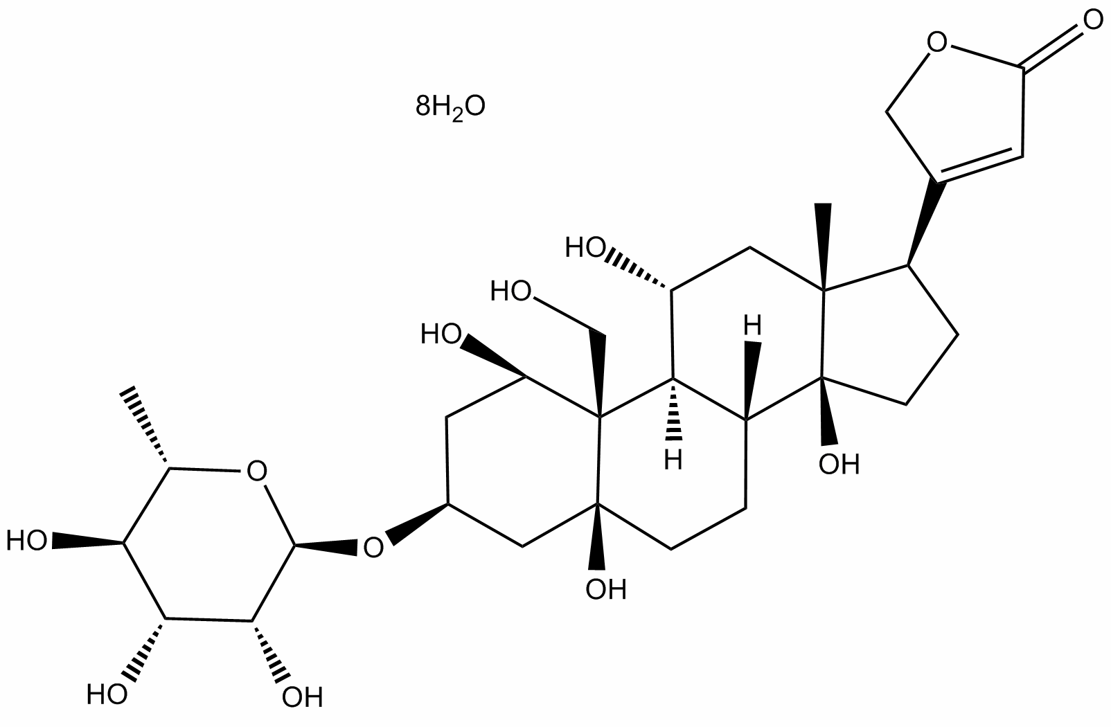 is regulated by PI3K and other factors. We found that thiamet-G treatment did not significantly alter the level of GSK-3b except after treatment for 9�C24 h, but blocked its phosphorylation at Ser9 completely, suggesting that GSK-3b was markedly activated under these conditions. Phosphorylation of GSK-3b at Tyr216, which makes it more active, was also increased 24 h after thiamet-G treatment. Consistent with the almost complete dephosphorylation of GSK-3b at Ser9, Ser473 and Thr308 phosphorylation of AKT, which determines its kinase activity, was also blocked by thiamet-G treatment. However, we did not find any significant changes of either the level or the activation of PI3K, the major upstream kinase of AKT, in the mouse brain after thiamet-G treatment. These results suggest that thiamet-G induced over-activation of GSK-3b via inhibition of AKT phosphorylation. The over-activation of GSK-3b may explain the increased tau phosphorylation at Ser199, Ser202, Ser396 and Ser422, NVP-BKM120 because these sites are the phosphorylation sites catalyzed mainly by GSK-3b. CDK5 is the second most important tau kinase in the brainand is activated by p35/p25. We thus also studied the level of CDK5 and its activators. We found that thiamet-G did not alter either CDK5 or p35. P25, which is the truncated and a more active form of p35, was not detectable in the mouse brain. To elucidate the complex regulation of site-specific phosphorylation induced by thiamet-G, we employed cultured cells, because cell cultures are more easily manipulated. We first selected AHP cells because they are more close to brain neurons than tumor cell lines. As expected, treatment of AHP cells with 20 nM thiamet-G increased protein O-GlcNAcylation. While we observed decreased tau phosphorylation at several phosphorylation sites in the AHP cells after thiamet-G treatment, it did not induce any significant increase in tau phosphorylation at the phosphorylation sites studied except at a transient elevation of Ser396 at 30 min after the treatment. We then investigated the levels and the activation of GSK-3b and the upstream PI3K-AKT signaling transduction pathway, as well as CDK5/p35. We did not find any significant changes upon the treatments with thiamet-G in AHP cells. These results further support our conclusion above that the increased tau phosphorylation at some sites observed in the thiamet-G treated mouse brains was due to GSK3b activation. To learn whether the phenomena we observed in undifferentiated AHP cells were specific to these cells, we also performed similar experiments in differentiated AHP cells and differentiated PC12 cells. As seen in proliferating AHP cells, we did not observe any marked elevation of tau phosphorylation at any phosphorylation sites or changes of tau kinases upon thiamet-G treatments in these two types of cells. Thiamet-G is a highly specific OGA inhibitor that was synthesized based on rationale design. Initial studies indicated that this compound reduce tau phosphorylation at some phosphorylation sites that can be abnormally phosphorylated in AD, suggesting that OGA inhibition may offer a potential therapeutic approach for slowing tau-mediated neurodegeneration seen in AD and other tauopathies. Because tau phosphorylation at different sites has different impacts on tau’s function and pathology, investigating the role of thiamet-G on site-specific tau phosphorylation is needed.
Monthly Archives: July 2019
The volume of water intake was measured daily to determine the amount of DSS consumed per mouse
Consistently, IFN-c production from CD4+ and CD8+ T cells in mesenteric lymph nodes was inhibited by bortezomib treatment. Thus, bortezomib treatment attenuated DSSinduced colitis by inhibiting excessive IFN-c production from CD4 + and CD8 + T cells. The immunoproteasome subunit 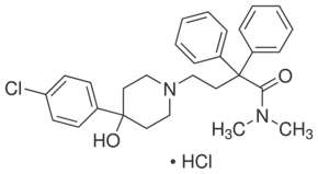 LMP7 is critical for proteasome activity, and it was recently reported that LMP7deficiency is associated with reduced severity of DSS-induced colitis in mice. Patients with inflammatory bowel disease exhibit high levels of LMP7 in the inflamed gut, and their increased proteasome activity induced by high levels of expression of immunoproteasome subunits mediates sustained activation of NF-kB. These results suggest that proteasome inhibitors may ameliorate inflammatory bowel disease. In this study, bortezomib administration substantially reduced the severity of DSS-induced colitis as well as significantly enhancing apoptosis and IkB expression of CD4 + and CD8 + T cells during DSS-induced colitis. Thus, NF-kB inhibition is likely to contribute to bortezomib-induced cell death in T cells, thereby suppressing DSS-induced colitis. Bortezomib treatment of mice with lupus-like disease significantly improves the disease severity by reducing the numbers of both CD4 + and CD8 + T cells in the spleen. Bortezomib treatment also demonstrates significant protection from acute graft-versus-host disease in a murine allogeneic bone marrow transplantation model by inhibiting allogeneic T cell proliferation. By contrast, bortezomib administration largely eliminates plasma cells but not T cells or B cells in murine models of human systemic lupus erythematosus. In the current study, bortezomib treatment reduced the numbers of CD4 + and CD8 + T cells, but not B cells or macrophages during DSS-induced colitis. It has been reported that proliferating T cells are more sensitive to Staurosporine bortezomibmediated cytotoxity than resting T cells. Proteasome inhibitors induce endoplasmic reticulum stress-induced apoptosis in multiple PCI-32765 myeloma cells as a result of the terminal unfolded protein response, while inhibition of proteasome activities by proteasome inhibitors induces apoptosis preferentially in rapid proliferating neoplastic cells. Thus, bortezomib treatment is likely to eliminate only excessively proliferating immune cells, thereby suppressing harmful inflammatory responses. Proteasome inhibition using bortezomib has recently emerged as an effective anticancer therapy. Thus far, the therapeutic feasibility of protease inhibition in inflammatory and autoimmune diseases has been revealed only in murine models of human systemic lupus erythematosus, experimental autoimmune encephalomyelitis, rheumatoid arthritis, asthma, and contact dermatitis. In the current study, bortezomib treatment in mice resulted in attenuated DSS-induced colitis, suggesting that bortezomib may also be effective for the treatment of human ulcerative colitis. Patients with this disease are generally treated with anti-inflammatory and immunosuppressive drugs, antizbiotics, and biologics such as anti-tumor necrosis factor therapies and/or surgery. However, such therapies do not cure the disease and patients suffer a life-long illness. Further studies are needed to determine the precise mechanisms by which bortezomib treatment reduces the severity of DSS-induced colitis. Nonetheless, if the efficacy seen in mice translates to humans, the current results may provide new insights and therapeutic approaches for treating ulcerative colitis.
The inhibitory potential of targeting two structurally distinct regions of the same protein may contribute to the synergistic effect
These findings are consistent with a recent study in melanoma cells in which dual treatment with the PI3K inhibitor PI-103 and rapamycin reversed compensatory Akt phosphorylation and induced cell cycle arrest, and xenograft studies demonstrated reduced tumor growth with this combination strategy. We extend these findings herein to define a potential mechanism by which the combination therapy promotes cell death. We found that BEZ235 alone blocked PI3K, mTORC1, and mTORC2 activity, in particular 4E-BP1 phosphorylation at a dose of 100 nM. However, BEZ235 was less effective in blocking rS6 phosphorylation. In comparison, temsirolimus completely abrogated phosphorylation of rS6 at 1 nM. Thus, combining both agentscompletely inhibited signaling throughout the pathway and synergistically induced cell death. Currently, combinatorial therapies are being applied to prevent resistance to single-agent treatments such as rapalogs. Examples of targeted small-molecule inhibitors under investigation include BEZ235, AZD2171; LBH589, LY294002, AZD6244, and ZSTK474. BEZ235 is a novel orally bioavailable inhibitor originally designed as a panPI3K family inhibitor based on the p110ckinase domain structure. Interestingly, when this compound was evaluated in preclinical studies, in vitro kinase assays revealed it also targets mTOR at a concentration of 20.7 nM. Therefore, BEZ235 is classified as a dual inhibitor that is capable of targeting both upstreamand downstreamof the PI3K/Akt/mTOR axis. BEZ235 has been reported to inhibit growth and proliferation and induce apoptosis in a variety of 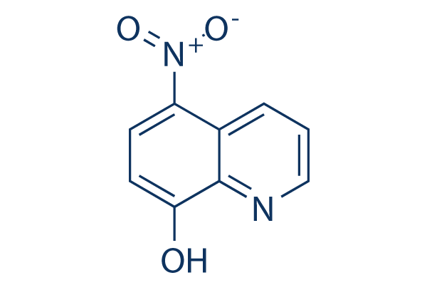 tumor cell lines, including breast cancer cells with mutant or amplified PIK3CA. BEZ235 showed antitumor activity in nude mice with few side effects. A recent report from a phase I study of BEZ235 in 59 patients with advanced solid tumors demonstrated antitumor effects and a favorable safety profile. ZSTK474, a pan-class I PI3K inhibitor, also demonstrated high potency against a panel of cancer cell lines and human tumor xenografts without toxicity to major organs. As discussed above, among all drugs tested, the agents which produced synergy with temsirolimus in our models were BEZ235 and ZSTK474. A main conclusion of our study is that combination treatment of ZSTK474 or BEZ235 with temsirolimus synergizes to decrease Y-27632 129830-38-2 viability in endometrial cancer cell lines. A potential mechanism of synergy from co-treatment with ZSTK474 and temsirolimus is the vertical NSC 136476 blockade of hyper-activated PI3K/Akt/mTOR signaling, specifically the simultaneous targeting of the upstream component PI3K by ZSTK474 and the downstream component mTORby temsirolimus. Temsirolimus alone only blocks rS6K activity downstream of mTORC1, whereas signaling through the other mTORC1 target 4E-BP1 is left intact. It has been documented in the literature that signaling through 4E-BP1 is required for Akt-mediated oncogenesis; therefore, inhibition of all components of this pathway is necessary to prevent tumor growth. Our data indicate that, in addition to inhibition of Akt activation, BEZ235 effectively blocks this residual signaling through 4E-BP1, which, when combined with temsirolimus inhibition of rS6K, synergistically blocks all arms of the PI3K/ Akt/mTOR pathway. Besides the observed inhibition of 4E-BP1 and rS6 with combined BEZ235and temsirolimus, another possibility might explain the observed synergy. Temsirolimus and BEZ235 target different structural domains of mTOR: temsirolimus is an allosteric inhibitor that targets the FKBP12-rapamycin-bindingdomain while BEZ235 is a catalytic inhibitor that targets the kinase domain.
PARP inhibition leads to apoptosis or senescence in cells where DNA repair by homologous recombination is impaired
For this study we used a potent selective PARP1-inhibitor Bortezomib 179324-69-7 BMN673, that has shown very encouraging results in phase I/II trials. Here we show that MRN is frequently lost in EC, which leads to increased PARP inhibitor sensitivity. This may be exploited for treatment of patients with EC harbouring loss of the MRNcomplex. The goal of this study is to show the frequency of loss of MRE11 and MRN-complex in EC and whether this leads to increased sensitivity to PARP-inhibitors exploiting MRE11 as a potential synthetic lethal gene. This is the first report to show that not only the protein expression of MRE11 but also the expression of the other members of the MRN-complex, RAD50 and NBS1, are lost in a substantial proportion of the ECs. Furthermore, we observed a significant association between protein loss of all MRN members as well as mismatch repair protein status in this large dataset. Protein expression of the MRN-complex has not yet been studied in EC tumours. In bladder cancer, MRE11 has been shown to exhibit predictive properties for radiotherapy treatment. Furthermore, complete loss of MRN-complex in MSI positive colorectal cancers is a frequent event, whereas loss of MRE11 in breast tumours is found only in 9% of cases. Although a clear correlation between mutational status and loss of protein expression could not be found in this study, it is remarkable that a high correlation of protein loss of all MRN members as well as MSI status was found in this large dataset. Confounding results on the mutational status of the MRE11 polyT allele may be related to known difficulties in sequencing of the MRE11 polyT allele due to mispairing of 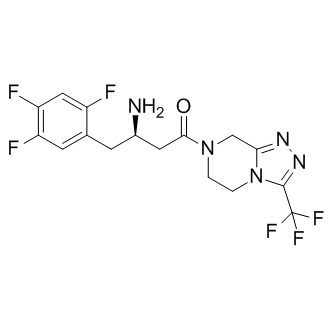 the polymerase enzyme. A previous study revealed high frequency of alterations of DSB repair genes in MSI positive ECs, where MRE11 and RAD50 exhibited heterozygous and homozygous mutations in 51% and 17%, respectively, without examining the impact of the loss of the proteins. Mutations of the MRE11 polyT allele are predominantly heterozygous mutations and it has been suggested that only those with mutations of two and more nucleotides as well as homozygous mutations have a functional impact in terms of loss of function. In this report, we cannot confirm the high frequency of intronic mutations but provide evidence that MRE11 protein is lost in a substantial proportion of ECs. Recently, it has been shown that whole exon sequencing of MRE11 revealed mutations in 1.9% of the EC tumours within the exons. However, intronic mutations have not been assessed, explaining why the frequency of MRE11 mutations is reported to be low not only in the study by Price et al. but also in a recent one by The Cancer Genome Atlas Research Network. PARP inhibitors have shown remarkable sensitivity in BRCA1/ 2-deficient tumour models in vitro as well as in clinical trials involving ALK5 Inhibitor II carriers of BRCA1/2 germ line mutations. Further evolving evidence, however, suggests the potential for a broader scope for PARP inhibitor activity. In fact, for EC we have previously proposed loss of PTEN expression as a potential biomarker for the treatment with PARP-inhibitors based on preclinical data as well as on a clinical case report. Other studies however, have been questioning the role of PTEN in HR, suggesting that this might be a cell line specific phenomenon. Nevertheless, the exact mechanism of the involvement of PTEN in HR DNA repair remains to be elucidated. Loss of MRE11 expression has been suggested to sensitize colorectal, breast and haematological cancer cell lines to PARP-inhibitors due to impaired HR DNA repair. Our report suggests, for the first time, the potential use of PARP inhibitors in the treatment of endometrial cancer based on preclinical findings.
The small molecule 10058-F4 has been extensively studied in the context of targeting c-MYC in cancer cells
As targeting its direct or indirect downstream targets. A number of small molecular compounds inhibiting c-MYC-MAX dimerization have been identified and among them 10058-F4 is by far the most studied. Biophysical experiments have shown that it interacts with the C-terminal bHLHZip region of c-MYC. A fluorescence polarization assay was used to determine the affinity as well as to identify the binding site of 10058-F4 on cMYC using different deletions, point mutations and synthetic peptides. NMR measurements confirmed that 10058-F4 binds to amino acid residues 402�C412 in the bHLHZip domain of c-MYC. Furthermore, metadynamic molecular simulations and an ion mobility mass spectrometry using a peptide corresponding to the identified binding site, indicated that the compound binds to an inactive, disordered conformation of cMYC. Together these studies suggest that 10058-F4 inhibits the function of c-MYC in a direct manner by preventing cMYC/MAX hetero-dimerization. Importantly, several reports have shown that 10058-F4 affects c-MYC expression and induces cell cycle arrest, inhibits cell growth, promotes apoptosis and confers chemo-sensitivity in a c-MYC specific manner in various cancer cell types. In addition, GSK2118436 Raf inhibitor treatment of acute myeloid leukemia cells with 10058-F4 leads to myeloid differentiation. The effect of 10058-F4 treatment in vivo has been investigated in xenograft models of prostate cancer but no significant antitumor activity could be observed, probably due to its rapid clearance and low potency. In contrast, we have recently demonstrated anti-tumorigenic effects of 10058-F4 in two tumor models of MYCN-amplified neuroblastoma, suggesting that direct MYC inhibition using a small molecule is achievable in vivo. The structurally unrelated small molecule 10074-G5 was identified simultaneously as 10058-F4 as another substance that inhibits the c-MYC/MAX interaction. This molecule also decreased c-MYC protein levels and inhibited cell growth, but failed to show any antitumor activity in a xenograft model using a Burkitt’s lymphoma cell line. The cognate binding site for 10074-G5 on c-MYC was found to be distinct from that of 10058-F4, spanning amino acid residues 363�C381. Both molecules were found to bind independently of each other, and probably induce only local conformation changes in the bHLHZip domain of c-MYC preventing its interaction with MAX. In order to identify more potent compounds, several analogs of 10058-F4 have been synthesized, some of which, including exhibited improved growth inhibition of c-MYC expressing cells. Since c-MYC and MYCN share structural similarity in the bHLHZip domain we hypothesized that 10058-F4 also targets MYCN. We have previously shown that this compound interferes with the MYCN/MAX interaction leading to cell cycle arrest, apoptosis, and neuronal differentiation in MYCN-overexpressing NB cell lines. In addition, using 10058-F4 as a tool, we found that inhibition of MYCN results in mitochondrial dysfunction leading to lipid accumulation. Importantly, 10058-F4 treatment furthermore increased the survival of TH-MYCN transgenic mice and showed anti-tumor effects in established aggressive NB xenografts. Here, we determined the direct binding of 10058-F4 and additional selected c-MYC-targeting compounds to MYCN by surface plasmon resonance. 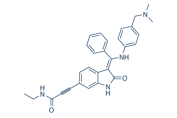 We found that all molecules previously reported to bind to c-MYC also bound to MYCN. Treatment with the small molecules furthermore interfered with the MYCN/ MAX interaction and caused protein degradation, apoptosis, differentiation and lipid formation to different extents in MYCNamplified NB cells. To date several research PLX4032 Raf inhibitor groups have focused on developing compounds that target c-MYC for cancer therapy, whilst there have been only a few publications that have explored the possibility of targeting MYCN.