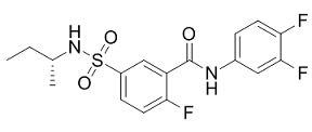
Because KORs are expressed in moderately high levels in the hippocampus, here we studied a potential role for hippocampal KORs in the renewal of extinguished fear. We found a dissociation in the contribution of hippocampal KORs, such that KORs in the VH, but not the DH, mediate the renewal of fear. The results from Experiment 1 showed that antagonizing KORs in the VH impaired the renewal of fear. Although recent research has clearly demonstrated a role for the VH in the return of fear  following extinction, the DH has also been shown to mediate renewal. Furthermore, the DH in the rat projects to the dorsal region of the mPFC, and contains KORs. These experiments investigated the role of hippocampal KORs in the renewal of extinguished fear. We demonstrated that: 1) intra-VH microinfusions of the KOR antagonist norBNI significantly attenuated renewal using a within-subjects design, 2) both a 5 mg and 10 mg dose were equally effective at reducing CSfreezing in the training context, and 3) intra-DH microinfusions of norBNI had no effect on the expression of renewal. Together these experiments reveal a dissociation in hippocampal contributions to renewal, where KORs in the VH, but not the DH, contribute to the renewal of extinguished fear. These findings are consistent with previous studies demonstrating involvement of the hippocampus in fear renewal. However unlike previous studies, here we show a clear dissociation in the contribution of distinct hippocampal regions. Prior functional studies investigating the neural circuitry underlying renewal have used lesions or temporary inactivation methods to target either the DH or VH. Such studies found that inactivating either hippocampal region stabilizing reproductive division labor maintaining link physiological state foraging behavior prevented renewal. Clearly both the DH and VH are essential components of the circuitry mediating the return of fear following extinction, yet here we demonstrate that the contributions of these regions are distinct. Specifically, we demonstrated that only the VH involvement in renewal relies on activation of KORs, at least in part. Recently, Orsini end colleagues demonstrated that the VH mediates renewal via direct projections to the mPFC and BA. This raises the question of how KORs within the VH are acting on this circuit to mediate renewal. One possibility is through the extensive projections from the VH to the BA. Renewal increases Fos expression in BA-projecting neurons in the VH, and results in increased firing in a population of BA neurons receiving input from the VH. This suggests that recruitment of this VH-BA pathway is activated during renewal. Although activation of KORs in the hippocampus has been shown to inhibit excitatory transmission, in regions of the caudal hippocampus KORs are also located on GABAergic interneurons. As such, the attenuation of renewal seen here in Experiment 1 is potentially due to norBNI acting on these GABAergic interneurons to prevent the disinhibition of pyramidal neurons, reducing activation of the VHBA pathway and thus diminishing the response of fear neurons in the BA. Recently however, it was demonstrated that individual VH neurons send convergent projections to both the BA and mPFC, including the prelimbic cortex. This is of note considering the role of the PL in renewal and the expression of conditioned fear. For example, the PL shows significant neuronal activation during renewal, and CS-evoked firing which correlates with learned freezing behavior. Such findings raise the possibility that the attenuation of renewal by infusions of norBNI into the VH was due to simultaneous reduction in activity in both PL and BA. Of course it is important to note that KORs are widely distributed in the hippocampus, including on granule cell mossy fibres and perforant path terminals, and hence could exert numerous effects on hippocampal neurons.
Monthly Archives: March 2019
Under investigation more than any other organelle due to their vulnerability to oxidative damage and their contribution to apoptosis
As a result of limited therapeutic accumulation within mitochondria, targeting the mitochondria with antioxidants or therapeutics has been a major interest especially for cardiovascular disease and cancer. Small molecules can permeate through the mitochondrial outer membrane but fail to cross the inner membrane. Taking advantage of the high inner membrane potential gradient, lipophilic cations can easily accumulate within the mitochondria as well as permeate the phospholipid bilayers. Vitamin E conjugated to TPP + can accumulate into the mitochondria, where it decreases ROS more effectively than vitamin E alone, and is able to ameliorate oxidative stress-mediated disease. While conjugating vitamin E to TPP + has been previously described, our goal was to conjugate the vitamin E metabolite, a-CEHC, to TPP+ and to design a fast and efficient synthetic method using a lysine linker and solid phase synthesis. This method does not require isolation of synthetic intermediates, while reagents and by-products are washed away after each step. In addition, similar to trolox, a-CEHC contains the a-tocopherol ring structure but have a truncated side chain with one carbon longer than trolox. The chroman ring of vitamin E becomes redox active at the mitochondria, where it forms semiquinone after detoxifying a free radical via hydrogen donation. The semiquinone is further reduced by intramitochondrial ascorbic acid or by electron donation. The chroman ring is still intact in aCEHC when conjugated to TPP +. A lysine linker with two protecting groups was used, which enabled the conjugation of TPP + and a-CEHC. The masked lysine was coupled onto the Rink Amide MBHA resin. HBTU and HOBt were used to enhance the coupling rate. The Fmoc was then deprotected to allow for TPP + conjugation through its carboxylic acid group forming an amide bond. The Mtt protecting group was then removed. The removal of the protecting group enabled the carboxylic acid on a-CEHC side chain to form an amide bond with the lysine linker. The final product, TPP + -Lysine-a-CEHC, was then released from the resin via treatment with 95% TFA. The final product was characterized by MALDI-TOF mass spectrometry. The molecular weight peak was at 736.39, which corresponds to the expected peak for the MitoCEHC generated by ChemDraw software. The mass spectrometry data also shows virtually no trace of by-products, reagents or synthetic intermediates. The ability of final product to diminish oxidative stress was examined in vitro. Oxidative stress is defined as the overproduction of oxidizing chemical species and the failure to eradicate their excess by enzymatic or non-enzymatic antioxidants. Elevation in ROS production is a factor in the etiology of cardiovascular disease by modifying lipids, proteins, and nucleic acids. To further explore the antioxidant activity of the conjugated MitoCEHC, the oxidation of CM-H2DCFDA was measured. The H2DCFDA derivative with a thiol-reactive chloromethyl group was used due to its better retention in live cells than H2DCFDA. This derivative is retained better in cells because of its ability  to bind covalently to intracellular components. BAEC were incubated with low and high glucose concentrations. The cells incubated under hyperglycemic conditions showed an increase in ROS production, which is mainly in the mitochondria. Flow cytometry data also showed decrease in ROS production in the hyperglycemic cells treated with MitoCEHC.
At http://www.neuroscienceres.com/index.php/2019/02/27/introduce-possibility-increases-mt-i-mt-ii-mrna-expression-decreases-total-zinc/, we supply the most recent news and developments about In the case of livestock species where no embryonic stem cell line with germ-line characteristic.
Our results indicate that DCP could be improved by in vitro electrical stimulation
Therefore, we speculated that it may be resulted from the increased activity of cholinergic receptors, the promoted release of Ach and the decreased expression of Ach. The underlying mechanism for the DCP improvement may be due to increases of cAMP in bladder, which could modulate the signaling pathways of neurotransmitter and receptors and increases of CGRP expression in bladder wall and DRG, leading to the enhancement of the contractility of the detrusor and sense of bladder filling. We have established a Rel report transgenic mouse model which can be used to monitor the endogenous Rel expression. Using in vivo bioluminescence imaging detection system, we demonstrated that luciferase activity in B6-Tg8Mlit mice was dramatically induced after i.p injection of LPS. The result of ex vivo experiment showed that the luciferase expression was induced in the heart, liver, spleen, lung, kidney, intestine, stomach, thymus and macrophages after the treatment with LPS, especially in the heart, liver, spleen, intestine and stomach. The data were in consistent with the change of endogenous murine Rel mRNA expression induced by LPS treatment. Meanwhile, the patterns of luciferase expression in LPS-treated B6-Tg8Mlit mice were also comparable to the results of Rel expression reported previously. The fold change did not match exactly between the luciferase activity and endogenous Rel mRNA expression. However, this is understandable that the protein level is often not in linear correlation with the endogenous mRNA expression in cells. Dexamethasone and aspirin, two well-known antiinflammatory drugs, suppressed the induction of luciferase expression and endogenous Rel expression in LPS treated B6-Tg8Mlit mice. These data demonstrate that the transgenic mice are faithful for monitoring Rel expression in vivo in c-Rel involved physiological or pathological processes and for evaluating the effects of anti-inflammatory drugs. LPS could induce Rel expression in monocytes and macrophages. We also collected macrophages from the abdomen fluid of the transgenic mouse. These specific cells manifested a good response of luciferase expression to LPS stimulus in vitro and could be used for high-throughput screening and studying of anti-inflammatory drugs at cellular level. Zymosan, a cell wall particle derived from Saccharomyces cerevisiae, is also widely used to induce inflammation in various relative experiments. However, the receptors for zymosan are distinct from those for LPS. LPS, the part of the outer cell wall of Gram negative bacteria, is detected by TLR4, while zymosan is recognized by TLR2 and TLR6. Although LPS and zymosan activates different triggers of target cells, the following inflammatory cascades seem to be similar. Our data also support the conclusion, for both LPS and zymosan could induce similar luciferase expression profiles in the B6-Tg8Mlit mice. The mouse EAE model is routinely used to study molecular mechanisms and signaling pathways of inflammatory regulation in Multiple sclerosis. It was reported that c-Rel-deficient mouse was resistant to the development of EAE due to its defective in the IL-12 and IFN-c induction and in the Th1 responses. It suggested that Rel expression was involved in the disease process of EAE. In our experiments, the luciferase expression in EAE group increased significantly at the eighth day after MOG injection before the loss of body weight and clinical symptoms occurred.
Have a look at http://www.proteintyrosinekinases.com/index.php/2019/02/22/formed-majority-sugars-oxidized-sugar-aldonic-acids/ for intriguing information on www.neuroscienceres.com.
Hormone signaling pathways play an important role in prostate cancer development
These findings, combined with a lack of significant correlation between leptin and appetite, suggest that leptin reductions in TB are a reflection of wasting seen in TB disease, rather than a driving force behind appetite and nutritional dysregulation. We found that ghrelin in TB patients is elevated compared to controls, falls with treatment, and correlates negatively with BMI and BF. Our findings conflict with the one prior study we found on ghrelin levels in TB, which reported no differences in baseline or post-treatment ghrelin concentrations in TB patients and reported lower ghrelin levels in malnourished cases compared to wellnourished cases. Our results do agree with studies examining ghrelin in other pulmonary disorders, which found elevated ghrelin in malnourished patients with COPD and lung cancer. While no other published studies have examined resistin in infections, our finding of elevated resistin in the disease state agreed with prior studies showing elevations in gastrointestinal cancers, direct correlations between resistin and cancer stage and resistin and BMI loss. In summary, our data show that patients with pulmonary TB display clear AbMole alpha-Cyperone alterations in energy regulatory hormones in comparison to healthy controls, and these alterations coincide with changes in appetite and nutritional status. As altered hormone levels normalized during treatment, appetite and nutritional status also improved. PYY was the strongest predictor of appetite in these patients and high PYY was an indicator of poor prognosis, with high levels predicting reduced gains in appetite and body fat during treatment. While previous studies have examined various combinations of energy-regulatory hormones in patients with TB, we are unaware of any studies which have evaluated PYY, leptin, ghrelin, and resistin in the same population, or any that have three longitudinal data points during treatment. This broad view provides valuable insight into the patterns of disrupted energy regulation and inflammation in TB. In addition, this was the first published study to examine PYY in TB and our results suggest this hormone is a key player in appetite and energy dysregulation in TB. The relatively short follow-up time of this study limited our ability to measure long-term correlations between hormones, appetite, and nutritional status during treatment. While we found strong correlation trends between PYY and appetite as well as BF, we did not detect a correlation between PYY and BMI gain, nor could we detect correlations between appetite and BMI/BF gain during treatment. BMI and BF likely lag behind appetite, with appetite improving first during treatment and weight gain happening as a result. Thus, a longer follow-up time may have demonstrated stronger correlations between initial PYY and appetite and weight changes during or following treatment. To rule out the possibility that changes in hormones reflect differences in body composition rather than the disease state itself, it would have been ideal to match cases and controls by BMI and BF. However, as TB generally causes cachexia, healthy subjects by nature do not have equivalent body composition to TB patients and thus BMI was not a feasible option to use as matching criteria. A future study comparing TB patients with those with other cachexia-inducing disease states could further explore the hormonal abnormalities specific to TB.
In addition airway hyperresponsiveness after methacholine challenge was more efficiency
The cytokine IL-6 regulates the functions of CD4 T cells and mediates asthma induction, whereas IL-12 regulates the Th1/Th2 balance and promotes IFN-c production. IFN-c is related to the persistence and severity of asthma. IL-4 and IL-13, which are key cytokines in the pathogenesis of asthma, are involved in airway remodeling, inflammatory processes, airway hyperresponsiveness, goblet-cell hyperplasia, eosinophil infiltration, mucus hypersecretion, and B cell activation. IL-5 regulates the development, activation, migration, and survival of eosinophils, which are characteristic features of asthma. Asthma is controlled with bronchodilators, corticosteroids, leukotriene modifiers, theophylline, and/or anti-IgE therapy; however, none of these treatments are curative. These data can be further utilized to establish novel clinical diagnostic tools to enhance subsequent clinical application Inhaled corticosteroids are commonly used, but in addition to their side effects, these drugs tend to reduce glucocorticoid receptorbinding affinity and T-cell response. Therefore, alternative therapies are sought from traditional medicines or other natural products that have therapeutic effects in respiratory disorders. In our study, we found that ACA dose-dependently suppressed WBC infiltration of the lungs in mice with OVA-induced asthma, and 50 mg/kg/day ACA treatment reduced the WBC count to that of the vehicle control group. Specifically, eosinophil infiltration, which is characteristic of asthma, was significantly suppressed by ACA. In addition, ACA blocked OVA-induced histopathological changes such as airway remodeling, goblet-cell hyperplasia, eosinophil infiltration, and mucus plugs. Although treatment with ACA did not inhibit B cell activation, as assessed by CD79a expression, our results show that ACA is effective at reducing populations of CD4+ Th cells and CD8+ cytotoxic T cells in the lungs of mice with OVA-induced asthma. Finally, ACA downregulated Th2 cytokines IL-4 and IL-13 and Th1 cytokines IL-12a and IFN-c, but did not affect the secretion of IL-5. The relationship between Th1 cells and Th2 cells plays an important role in the pathogenesis of asthma. Mamessier and Magnan hypothesized that there are three situations related to asthma. In a healthy subject, activation of Th1 and Th2 cells is balanced, and the level of regulatory T-cell activation is relatively low. In well-controlled asthma, the level of Th1 cell activation is similar to that of regulatory T cells, but Th2 cell activation is suppressed. In uncontrolled asthma, the level of Th2 cell activation is lower than that of Th1 cells, which in turn is lower than that of regulatory T cells. Thus, not only is the balance between Th1 and Th2 cells important, equilibrium is needed between Th1/ Th2 cells and regulatory T cells. The Th2 cytokines IL-4 and IL-13 promote acute inflammatory processes in the pathogenesis of asthma and structural changes in the airways;. We found that ACA dose-dependently reduced IL-4 and IL-13 levels in the lungs. In addition, ACA decreased IL-12 a and INF-c levels as effectively as dexamethasone. Asthma was traditionally though to be initiated by an imbalance between Th1 and Th2 cells, The functions of IL-12 have been fairly well characterized; however, the role of INF-c in asthma has been controversial. Although Caenorhabditis elegans extract was reported to ameliorate asthma symptoms by increasing INF-c expression, hydrocortisone, which is used to treat asthma, has been shown to decrease INF-c expression. Previous studies have reported elevated INF-c levels in the BALF and bronchioles of asthma patients.